

Why All on 4 Implants Can Transform a Full Smile
All on 4 implants replace a full upper or lower arch of missing or failing teeth with one fixed bridge supported by four strategically placed dental implants. Two front implants are usually placed straight, while the back two are tilted to use available bone, avoid key anatomy, and help reduce the need for extensive grafting.
For many people, this approach can offer a stable alternative to loose dentures. It may improve chewing, speech, comfort, and confidence, often with a temporary fixed set of teeth placed soon after surgery when implant stability and health factors allow.
All on 4 treatment is not one-size-fits-all. A safe plan starts with a detailed health review and 3D imaging to assess bone, gum health, bite, medications, and goals. At AZ Dentist Scottsdale, AZ Dentist Glendale, AZ Dentist Sun City, and AZ Dentist Sedona, the focus should always be on a plan that feels clear, comfortable, and right for you.
I’m Dr. Janne Lynch, and my background in cosmetic dentistry and oral surgery helps me plan All on 4 implants with both function and natural-looking results in mind. I also understand dental anxiety, so the next steps should feel organized, gentle, and easy to understand.
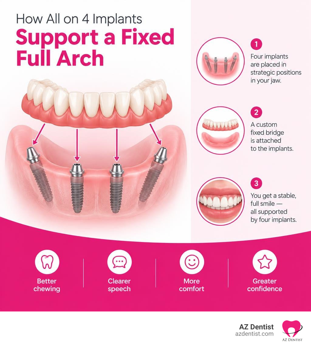
All on 4 implants terminology:
How All on 4 Implants Restore Full Arch Function
Full-arch rehabilitation fundamentally changes how bite forces travel through the jaw. In a standard dental arch, natural tooth roots distribute masticatory load directly into alveolar bone. When multiple teeth are lost, that stimulation vanishes, and bone begins to resorb.
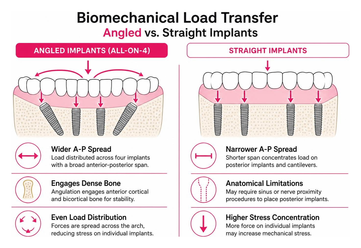
The biomechanical framework of full-arch therapy relies on cross-arch stabilization. By securing a rigid prosthetic bridge across four titanium or zirconia posts, occlusal forces spread evenly across the arch rather than concentrating on individual, vulnerable sites. A scientific review on the All-on-Four concept (Soto-Penaloza et al., 2017) confirms that splinting implants together with a rigid framework drastically decreases micromovement during the crucial early healing phase.
Learning all about full arch dental implants means understanding how modern engineering works alongside biological bone healing to deliver dependable biting strength.
Biomechanical Benefits of Angled Fixtures in All on 4 Implants
The hallmark innovation of this protocol lies in tilting the two posterior fixtures between 17 and 35 degrees. Rather than placing every post straight up and down, angling the rear implants offers critical clinical advantages:
- Anatomical Avoidance: Tilting allows fixtures to bypass the maxillary sinuses in the upper jaw and the mental foramen (and inferior alveolar nerve canal) in the lower jaw without requiring invasive relocation procedures.
- Cantilever Reduction: By exiting the bone further back along the dental ridge, angled fixtures widen the anterior-posterior (A-P) spread. This minimizes the length of unsupported bridge extensions (cantilevers), protecting the prosthesis against bending forces and fracture.
- Cortical Bone Anchorage: Angulation lets the surgeon engage dense anterior cortical bone and bicortical structures, maximizing primary stability even in an atrophic ridge.
Comparing Fixed Arches to Removable Alternatives
Many patients explore missing tooth replacement choices after years of struggling with shifting acrylic plates and messy adhesives. Traditional dentures rest on soft gum tissue, leading to continuous bone resorption underneath.
| Feature | All-on-4 Fixed Implant Bridge | Traditional Removable Denture |
|---|---|---|
| Stability | Screwed securely onto implants; zero slipping | Held by suction, muscle control, or adhesive pastes |
| Biting Efficiency | Restores 80% to 95% of natural chewing force | Delivers roughly 20% to 30% of natural chewing force |
| Palatal Coverage | Horseshoe design; roof of the mouth remains completely open | Full acrylic plate covers the hard palate, dampening taste |
| Bone Preservation | Stimulates alveolar bone, reducing long-term resorption | Accelerates ridge resorption due to surface pressure |
| Daily Maintenance | Brushed and water-flossed just like natural teeth | Must be removed nightly for soaking and separate cleaning |
| Speech Clarity | Minimal speech adaptation; low bulk profile | May cause clicking, slipping, or phonetic interference |
Comprehensive Assessment and Radiographic Evaluation
Every successful smile restoration starts with a meticulous preoperative workup. During initial diagnostic visits, we capture high-resolution clinical photographs, take digital intraoral scans, and generate diagnostic wax-ups to analyze facial aesthetics, smile line height, lip support, and vertical dimension of occlusion (VDO). Careful diagnostic planning like this helps identify ideal candidates for All-on-4 treatment and supports predictable, long-term surgical outcomes.
Careful clinical screening matters because the alveolar ridge must be evaluated not just for width and height, but also to confirm whether a patient truly needs a bone graft. Consulting our guide to understanding bone graft necessity in implants helps clarify how tilting posterior implants frequently bypasses bone grafting altogether.
Evaluating Medical Candidacy and Bone Density for All on 4 Implants
Systemic health dictates implant integration far more than chronological age. Key medical considerations include:
- Age and Vitality: Clinical meta-analyses reveal that patients aged 75 and older achieve implant survival rates comparable to, and sometimes exceeding, younger demographics (over 96%), proving age alone is not a disqualifier.
- Antiresorptive Medications: Patients taking standard oral bisphosphonates for osteoporosis carry a very low risk of Medication-Related Osteonecrosis of the Jaw (MRONJ) (roughly 0.001% to 0.01%). In contrast, high-dose intravenous antiresorptive therapies for oncology require strict multidisciplinary coordination.
- Glycemic Control: Well-managed type 2 diabetes with stable HbA1c levels is fully compatible with successful osseointegration.
- Radiation History: Prior head and neck radiation therapy below 45 Gy shows minimal added implant failure risk, whereas doses exceeding 60 Gy necessitate thorough hyperbaric and oncologic review.
- Extreme Maxillary Atrophy: When the upper jaw exhibits severe resorption that rules out standard tilted implants, zygomatic fixtures anchored directly into the malar bone offer an alternative with a 96.2% long-term survival rate.
Radiographic Planning and Computer-Guided Surgery
Three-dimensional Cone Beam Computed Tomography (3D CBCT) forms the foundation of modern implantology. Using multiplanar reconstruction, we visualize bone density in Hounsfield-equivalent units, map nerve pathways, trace sinus cavities, and measure available bone down to fractions of a millimeter.
We use CAD/CAM surgical guides to translate digital treatment plans into real-world surgery. Virtual planning software lets us set implant trajectories, target subcrestal placement depths, and choose custom step-drilling sequences before ever entering the operatory.
Surgical Protocols, Medications, and Emergency Management
On the day of surgery, treatment proceeds through clear, highly controlled stages:
- Sedation and Local Anesthesia: Ensuring absolute comfort through oral or IV conscious sedation combined with long-acting local anesthetic blocks.
- Flap Reflection and Alveolectomy: Raising a full-thickness mucoperiosteal flap and performing conservative bone leveling to create a smooth, flat prosthetic platform and hide the transition line behind the patient’s smile.
- Guided Osteotomy and Placement: Preparing osteotomy sites using incremental step drills under continuous chilled saline irrigation to avoid thermal trauma, followed by placing fixtures to achieve an insertion torque between 30 and 50 Ncm.
- Multi-Unit Abutment (MUA) Attachment: Affixing straight abutments on anterior implants and 17- to 35-degree angled MUAs on posterior implants to establish a level, parallel plane for the fixed bridge.
Pharmacological Protocols and Pain Management
Effective pharmacology ensures patient safety, minimizes swelling, and prevents infection:
- Prophylactic Antibiotics: Amoxicillin (2 g orally one hour preoperatively, followed by 500 mg three times daily for 5 to 7 days) or Clindamycin (600 mg preoperatively, followed by 300 mg four times daily) for penicillin-allergic patients.
- Anti-Inflammatory Corticosteroids: Preoperative or intraoperative administration of Dexamethasone (4 to 8 mg IV or orally) to reduce secondary tissue edema, trismus, and post-surgical bruising.
- Analgesic Regimen: A multimodal pain management strategy pairing Ibuprofen (600–800 mg every 6 hours) with Acetaminophen (500–1000 mg every 6 hours) to control inflammation and discomfort without excessive opioid reliance.
- Antiseptic Rinses: Chlorhexidine gluconate (0.12%) oral rinse twice daily starting 24 hours post-surgery to maintain low bacterial counts while surgical sites heal.
Managing Surgical Complications and Intraoperative Emergencies
Surgical teams must stay prepared to handle complications and medical emergencies promptly:
- Vasovagal Syncope: Managed immediately by placing the patient in the Trendelenburg position (legs elevated above heart level), ensuring an open airway, administering supplemental oxygen, and applying cold compresses.
- Local Anesthetic Systemic Toxicity (LAST): Monitored through continuous pulse oximetry and blood pressure tracking; managed by terminating local anesthetic delivery, managing the airway, and having 20% lipid emulsion ready for severe cases.
- Hemorrhage Control: Managed using localized pressure, electrocautery, bone wax, or hemostatic gelatin sponges in areas near nutrient canals.
- Biological Complications: Mitigating peri-implant mucositis and peri-implantitis through precise passive bridge seating, smooth prosthetic emergence profiles, and scheduled maintenance cleanings.
Prosthetic Workflows and Healing Timeline
Restoring a full arch involves distinct surgical and restorative steps:
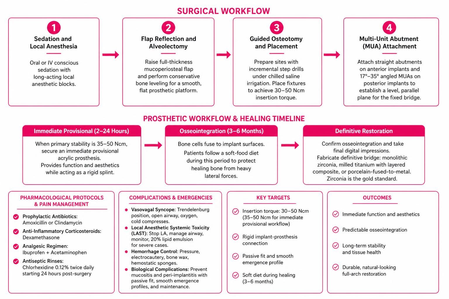
When primary stability meets or exceeds target torque thresholds (35–50 Ncm), an immediate provisional acrylic prosthesis is secured within 2 to 24 hours. This provisional bridge provides functional chewing and aesthetics while acting as a rigid splint.
Over the next 3 to 6 months, osseointegration occurs as living bone cells fuse to the titanium implant surfaces. Patients stay on a soft-food diet during this window to protect the healing bone from heavy lateral chewing forces.
Once osseointegration is confirmed, we take final digital impressions to fabricate the definitive bridge. Patients can explore All-on-4 alternatives and benefits—including monolithic zirconia arches, milled titanium frameworks with layered composite, or porcelain-fused-to-metal bridges. Zirconia remains the gold standard for full-arch restorations due to its high fracture resistance, smooth tissue surface, and lifelike translucency.
Frequently Asked Questions About All-on-4 Treatment
What is the long-term success rate of full-arch implants?
Long-term clinical studies report cumulative survival rates exceeding 99.8% at two years, with 10-year success rates holding steady between 94.8% and 98% (Soto-Penaloza et al., 2017). Because the bridge is supported by multiple integrated fixtures, the system remains exceptionally stable over decades when maintained with routine cleanings.
Can I receive implants if I have significant bone loss?
Yes. The All-on-4 protocol was specifically engineered for patients with severe bone atrophy. By angling the rear implants, we maximize contact with the dense bone at the front of the jaw and avoid anatomical boundaries like the maxillary sinus. While some complex cases benefit from reviewing a detailed guide to bone grafts, most full-arch patients can proceed without invasive bone grafting.
How long is the recovery period following surgery?
Most patients resume light daily activities within 2 to 4 days. Mild swelling and bruising typically peak around day three and resolve by the end of the first week. While the immediate provisional bridge lets you smile and speak normally right away, you should stick to a soft-food diet (eggs, fish, pasta, smoothies) for the first 3 to 4 months while the implants fully integrate with the jawbone.
Conclusion
Full-arch rehabilitation with All-on-4 implants offers a reliable way to replace failing or missing teeth, restore full chewing ability, preserve facial structure, and bring back a natural smile.
Whether you visit us at AZ Dentist Scottsdale, AZ Dentist Glendale, AZ Dentist Sun City, or AZ Dentist Sedona, our priority is to deliver comfortable, compassionate, and precise care tailored to your health goals. We believe in taking the time to listen, plan every detail using modern 3D imaging, and ensure you feel confident at every step of your journey.
If you are ready to explore your options, schedule a personalized consultation for comprehensive dental care at AZ Dentist and take the first step toward the smile of a lifetime.
References
Soto-Penaloza D, Zaragozí-Alonso R, Penarrocha-Diago M, Penarrocha-Diago M. “The all-on-four treatment concept: Systematic review..” Journal of clinical and experimental dentistry, 2017. PMCID PMC5347302.